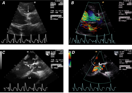
Fig. 3 Transthoracic echocardiograms (parasternal long-axis views) of aortic valve endocarditis show A ) the aortic valve, which is thickened and distorted by the vegetation (arrow), and B ) severe aortic regurgitation (arrow), in color-flow Doppler. Transesophageal echocardiograms (intraoperative midesophageal views) of the aortic valve show C ) a clear subvalvular vegetation (Veg) and the suggestion of perforation (Perf), and D ) findings that appear to be consistent with a perforation of the leaflet (arrow), in color-flow Doppler. Ao = aorta; LA = left atrium; LV = left ventricle

An official website of the United States government
Here's how you know
Official websites use .gov
A
.gov website belongs to an official
government organization in the United States.
Secure .gov websites use HTTPS
A lock (
) or https:// means you've safely
connected to the .gov website. Share sensitive
information only on official, secure websites.
