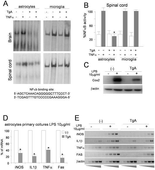
Figure 2. Inhibition of NF-κB activity in astrocytes from GFAP-IκBαAA mice.
(A) EMSA of nuclear protein extracts (5 µg) from primary astrocytes and microglia from brain and spinal cord of transgenic mice line A (Tg-A) and non transgenic mice (-) stimulated or unstimulated for 20 min with recombinant TNFα (10 ng/ml) before analysis. For NF-κB consensus probe see underlined sequence below. (B) Quantification of the experiments in (A) performed with spinal cord astrocytes and microglia by densitometric assay. Results obtained from non transgenic stimulated cells were defined as 100%. Results are expressed as the means +/− SEM of three independent experiments. *P<0,01. Effect of 3 h LPS treatment (10 µg/ml) on different NF-κB controlled gene in primary astrocytes from non transgenic (-) or GFAP-IκBαAA (TgA) transgenic mice. (C) Western blot analysis on protein extract using antibodies against COX2 and β-actin. (D) semi-quantitave PCR for iNOS, IL-1β, TNFα, Fas mRNA expression. Results obtained with cells from stimulated non transgenic mice are defined as 100% and compared with their corresponding results from stimulated transgenic samples. Data are normalized to β-actin. *p<0.05 **p<0.01. Results represent the mean +/−SEM of three groups. (E) Representative semi-quantitave PCR performed on cDNA from total RNA extracted from astrocytes primary cultures from non transgenic (-) and transgenic (Tg-A) mice untreated and treated with LPS 10 µg/ml.

