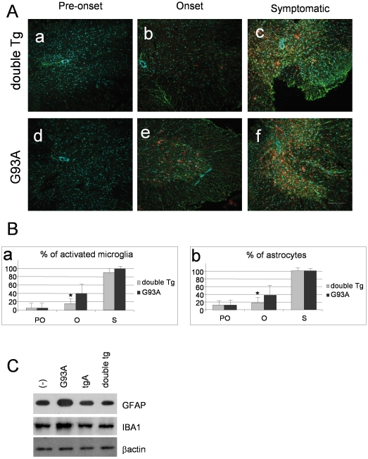
Figure 5. Selective expression of IκBαAA in astrocytes causes a weakly delayed microgliosis and astrocytosis only at the onset of disease (120 days).
(A) Immunofluorescence against GFAP (green) and Cd11b (red) in sections of lumbar spinal cord from 100 days (pre-onset), 120 days (onset) and 140 days (symptomatic) old double transgenic mice GFAP-IκBαAA/SOD1G93A (a,b,c) and SOD1G93A mice (d,e,f). Scale bar 100 µm. (B). Schematic representation of counting results in (A). Data are expressed as percentage (mean+/−SEM) of activated microglia (a) or astrocytes (b) from 3 different animals from the three different disease stages: pre-onset (PO); onset (O); symptomatic (S). The number of cells recorded from SOD1G93A mice at the symptomatic stage was considered 100%. *p<0.05. (C) GFAP and IBA1 protein levels were evaluated by Western blot on 20 µg of proteins/sample. Blots were probed for β-actin as a control.

