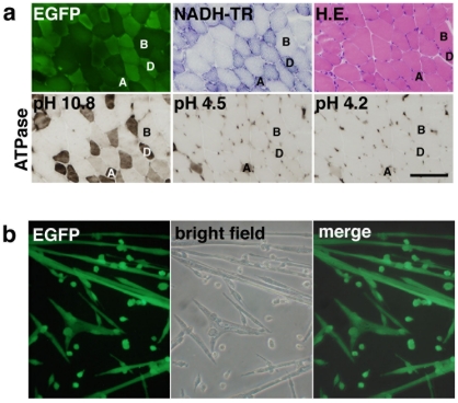
Figure 2. Intramuscular injection of LvMSCV-EGFP into striated muscle.
(a) Micrographs of serial sections from a lentiviral vector-transduced skeletal muscle showing fluorescent EGFP expression and NADH-TR, HE, and ATPase staining (at pHs 10.8, 4.5, and 4.2). EGFP expression was strong in the Type IID/X fibers (D), which can be distinguished by dark staining with NADH-TR and ATPase at pH 10.8, but weaker in type IIB fibers (B), which are lightly stained in NADH-TR. There are a few type IIA fibers (A), darkly stained with NADH-TR and ATPase at pHs 10.8, 4.5, and 4.2, also brightly positive for EGFP. Scale bar represents 100 µm. (b) Primary cultured myotubes isolated from LvMSCV-EGFP-injected TA muscles. Seven weeks after transduction, muscle cells were isolated and cultured in F10C/15% HS/5 ng/ml with bFGF. EGFP-positive myotubes were observed after 7 days in culture.

