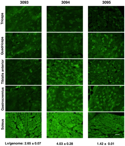
Figure 4. EGFP expression regulated by MSCV promoter in skeletal muscles following embryo transduction.
Brachio-triceps, quadriceps, tibialis anterior, gastrocnemius, and soleus muscles of 3 different mice (3093, 3094, and 3095) from independent embryo-transductions were cryosectioned and observed under a fluorescence microscope. The average Lv-copy numbers (mean ± SEM) are shown at the bottom. Each section shows the expression of EGFP. Although the overall levels of EGFP expression varied among the three mice (transduction at the two-cell embryo stage), they each displayed a similar fiber-type expression pattern. Scale bar represents 100 µm.

