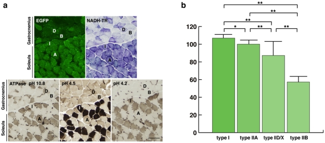
Figure 5. MSCV-EGFP transgenic mice showed fiber type-dependent variations in EGFP protein expression.
(a) The expression of EGFP at the border of the gastrocnemius and soleus muscles, where all 4 fiber types, type I, type IIA, type IIB, and type IID/X (labeled I, A, B, and D, respectively), are present. Each fiber type was confirmed by NADH-TR and ATPase (at pHs 10.8, 4.5, and 4.2) staining. Bar represents 100 µm. (b) Bar graph showing the mean (+SEM) intensity levels of EGFP protein in each fiber type. Type I fibers and IIA fibers presented the strongest signals, type IID/X fibers had intermediate expression levels, and type IIB fibers had the weakest. Except between type I and type IIA (*p = 0.03), there were significant differences among the various fiber types (**p<0.005).

