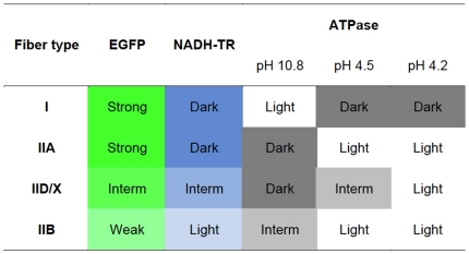
Figure 6. Summary of the fiber type-dependent EGFP expression in LvMSCV-EGFP mediated transgenic mice.
Levels of EGFP expression and typical staining pattern of each muscle fiber type are summarized. The EGFP expression pattern was mostly identical to that of the NADH-TR staining. Fiber types were determined by the ATPase stainings with different pHs (pH 10.8, 4.5, and 4.2). Colors reflect the staining colors on muscle sections.

