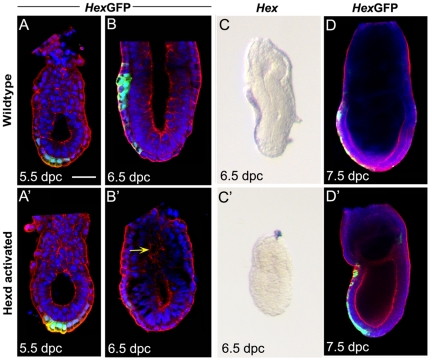
Figure 2. Ablation of the Hex-AVE in Hexdact/+ embryos.
(A–D) Hex-GFP and Hex expression in wild-type and Hexdact/+ embryos. (A–A′) At 5.5dpc n = 1/7 Hexdact/+ embryos have lost expression. Green arrow points to Hex-GFP domain of AVE; (B–B′) at 6.5dpc n = 4/12 have severely reduced or lost expression, yellow arrow indicating disorganised pro-amniotic cavity; (C–C′) Hex transcript is reduced or lost in n = 6/6 Hexdact/+ embryos at 6.5dpc, but (D) not affected at 7.5dpc in these embryos, n = 0/4. Scale bar 40 µm in A–A″; 60 µm in B; 90 µm in C and 100 µm in D. GFP (green) Nuclear stain (blue); F-actin (red).

