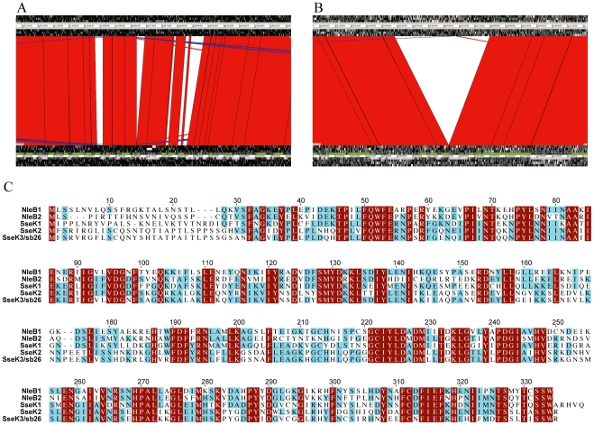
Figure 1. Phage encoded genomic differences between S. typhimurium SL1344 and LT2.
A & B. Screenshots from ARTEMIS COMPARISON TOOL showing (A) differences in the SopEϕ/Fels-2 region and (B) the ST64B insertion in SL1344 relative to LT2. The SL1344 sequence is shown on top and LT2 at the bottom. The red regions indicate alignment between the two genome sequences and the white regions indicate gaps in the alignment. C. Alignment of the amino acid sequences of SseK3 and other SseK/NleB family members. Residues conserved in all five proteins are shaded in maroon, those conserved in three or four proteins are shaded in cyan. SseK1 and 2 sequences are from LT2 and NleB1 and 2 are from E. coli strain EDL933.

