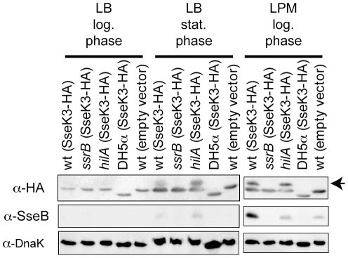
Figure 2. Expression of SseK3 in culture medium.
Western blots of whole bacterial cell lysates from the culture conditions and strain genotypes indicated at the top of the figure were probed with the antibodies indicated to the left of the respective panels. The anti-HA panels show a cross-reacting band in each lane just below the SseK3 band. This cross-reacting band is lower in the E. coli lysates than the Salmonella lysates. The arrow to the right of the α-HA panel indicates the bands corresponding to SseK3-HA.

