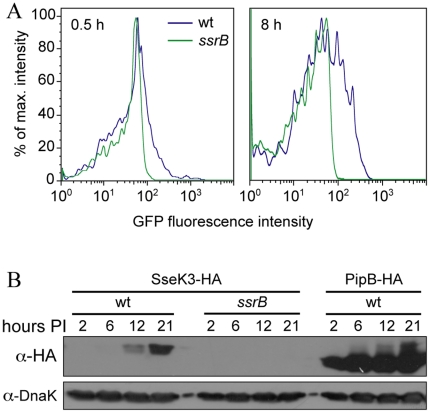
Figure 3. Expression of SseK3 in host cells.
A. Graphs showing GFP fluorescence intensity of intracellular wild-type or ssrB Salmonella containing an sseK3 promoter GFP fusion plasmid. Cells were infected for 0.5 h and 8 h before being processed for flow cytometry. B. Western blots of infected HeLa cell lysates made at the indicated times post-infection (hours PI), with the indicated strains transformed with plasmids encoding the indicated effector fused to tandem HA epitopes. The western blots were probed for the HA epitope and DnaK as a loading control.

