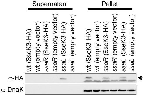
Figure 4. Secretion of SseK3.
Western blots of concentrated culture supernatant proteins and whole Salmonella cell lysates (pellet) from strains genotypes indicated at the top of the figure were probed with the antibodies indicated to the left of the figure. The arrow to the right of the α-HA panel indicates the bands corresponding to SseK3-HA.

