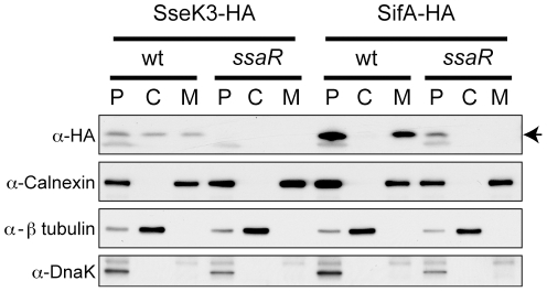
Figure 5. Translocation of SseK3 into infected HeLa cells.
Western blots from fractionated, infected HeLa cells probed with the antibodies indicated to the left of the figure. SseK3-HA and SifA-HA indicated at the top of the figure refer to the HA-tagged protein expressed in the respective strain, and under this, wt and ssaR refer to the genotype of the respective strain. P, C and M refer to the fractions collected from the infected cells as described in the text. The arrow to the right of the α-HA panel indicates the bands corresponding to SseK3-HA and SifA-HA (which are approximately the same MW).

