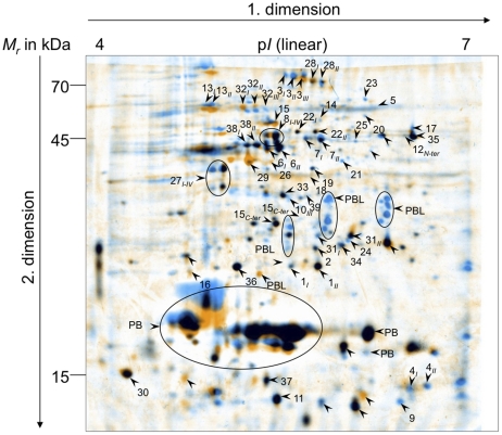
Figure 1. Proteomic comparison of Microcystis aeruginosa PCC 7806 wild type and microcystin-deficient ΔmcyB mutant.
Proteome of soluble fraction of Microcystis aeruginosa PCC 7806 wild type (WT, blue colouring) and microcystin-deficient ΔmcyB mutant (orange colouring) after exposition to high light of 70 µmol photons m−2s−1 for 2 hours. Gels were obtained after separation of soluble protein extracts (400 µg) by 2-DE (first dimension: IEF pH range 4–7 linear, second dimension: SDS-PAGE applying 12.5% acrylamide) and Coomassie-staining. Representative well resolved gels of WT and ΔmcyB were grouped, respectively, warped and fused to one gel via Delta2D version 4.0 (DECODON, Greifswald, Germany). Differential protein spots and selected non-differential protein spots analyzed for standardization are indicated with arrows. Highly abundant phycobiliproteins (PB), associated linker proteins (PBL) as well as proteins of more than three isoforms are encircled. Protein identities are given in Table 1 and Table S1.

