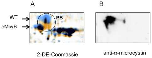
Figure 3. Comparative proteomic analysis of phycobiliproteins.
A) Mass shift of phycobiliproteins (PB) in Microcystis aeruginosa PCC 7806 wild type (blue colouring) compared to microcystin-deficient ΔmcyB mutant (orange colouring) as seen by 2DE-analysis. Proteins with differential mass in wild type and mutant are encircled. B) Corresponding immunoblot analysis using the microcystin-specific antibody.

