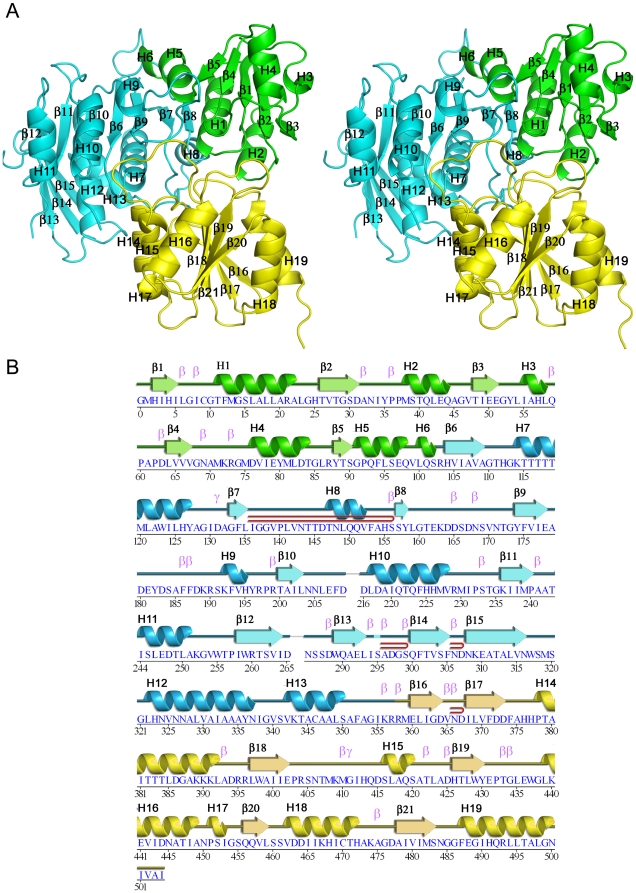
Figure 3. Crystal structure of PaMpl.
(A) Stereo ribbon diagram of PaMpl highlighting the 3 domains: N-terminal domain (ND), green; Middle Domain (MD), cyan; and C-terminal Domain (CD), yellow. (B) Diagram showing the secondary structure elements of PaMpl colored by domain and superimposed onto its primary sequence, adapted from PDBsum (http://www.ebi.ac.uk/pdbsum), where α-helices and 310-helices are sequentially labeled (H1, H2, H3 etc), β-strands are labeled (β1, β2, β3, etc), β-hairpins are indicated by red loops, and β- and γ-turns are indicated as β and γ.

