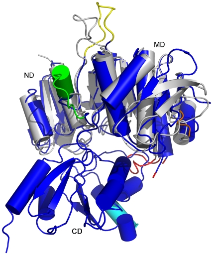
Figure 4. Comparison of crystal structures of full-length PaMpl and truncated NmMpl.
The crystal structure of truncated NmMpl (grey, ND and MD only, PDB id 3eag) is similar to PaMpl (blue) and can be superimposed with an r.m.s.d. of 2.2 Å over 311 Cα atoms with a sequence identity of 57%. The main differences are: NmMpl lacks the PaMpl-specific sequence segments 141–152 (green), 262–285 (orange) and 444–450 (cyan); NmMpl residues 152–163 are positioned differently compared to the corresponding residues 161–171 in PaMpl (yellow); and the segment corresponding to a loop (residues 199–208, red) in NmMpl is disordered in PaMpl (residues 210–215, flanking residues 209 and 216 are in red).

