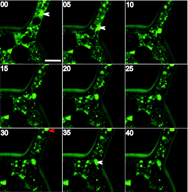
Figure 4.
Time-lapse series on AMF live spores. Panels represent 5 min intervals in a time-lapse sequence monitoring nuclear migration into a developing spore of G. diaphanum by confocal microscopy. SytoGreen stained nuclei move unidirectionally into the spore. Images were acquired on xyt mode. Arrowheads indicate the first (white) and second (red) nucleus entering the spore. Note that some nuclei are out of focus in the xy optical section and can be difficult to visualize. Scale bar on the top-left panel represents 10 μm. Numbers on the top left of each panel are minutes.

