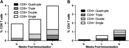
Figure 4.
IFN-γ, IL-2, TNF-α, and CD107a multifunctional cells detected by ICS in CD3+CD8+ and CD3+CD4+ populations. Mean percentage of quadruple (black), triple (dark gray), double (light gray), and single (white) functional cells detected within the CD8+ (A) and CD4+ (B) populations. Within the CD8+ population, the most frequently detected triple positive cells were CD107a+IFN-γ+TNF-α+; the most frequently detected double positive cells were CD107a+TNF-α+; and CD107a+ cells were the most frequently detected single positive cells. Within the CD4+ cells, the most frequently detected triple positive cells were CD107a+IFN-γ+IL-2+; the most frequently detected double positive cells were IFN-γ+IL-2+; and TNF-α+ cells were the most frequently detected single positive cells. At all time points the frequency of antigen-specific cytokine positive cells was greater in the CD8+ population (week 0, CD8+ = 1.92% and CD4+ = .12%; week 1, CD8+ = 2.43% and CD4+ = .58%; week 8, CD8+ = 3.96% and CD4+ = 1.19%).

