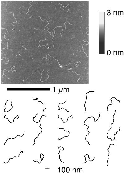
Figure 1.
(Upper) An example tapping-mode SFM image of DNA palindromic dimers deposited on a surface of freshly cleaved ruby mica. The height of the features on the surface are coded in shades of gray according to the color bar. (Lower) A subset of the molecular profiles digitized from the SFM images and used for the curvature analysis are shown.

