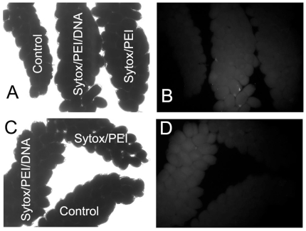
Figure 1. DNA uptake in developing oocytes.
(A) Under the optical light under 40X magnification. (B) The same image as in panel A but under the fluorescent light. (C) Under the optical light under 40X magnification. (D) The same image as in panel C but under the fluorescent light. Females were microinjected with 0.5 µl of Sytox/PEI/DNA or Sytox/PEI complex at 18 hours post bloodmeal (PBM) or without injection (control). The ovaries were dissected out at 42 hours PBM and observed under the fluorescent microscope (Nikon DiaPhot, Nikon Inc., Melville, NY).

