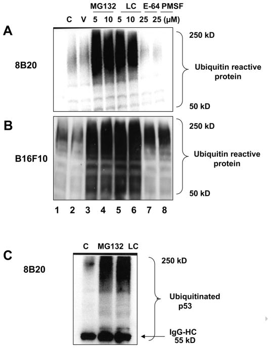
Figure 2.
(A, B) Treatment of murine melanoma cells with proteosomal inhibitors MG132 and clasto-lactacystin-β-lactone results in the generation of high molecular weight ubiquitin reactive products Exponentially growing 8B20 and B16F10 mouse melanoma cells were either untreated (C: control) or treated with ethanol (V: vehicle control), proteosomal inhibitors (MG132 and LC: clasto-lactacystin-β-lactone) or non-proteosomal inhibitors (E-64 and PMSF) at the indicated concentrations for 6 hours. Cell lysate (100 μg) was loaded on a 12 % polyacrylamide gel and subjected to SDS-PAGE and immunoblot analysis. Membranes were probed with the anti-ubiquitin antibody. (C) Detection of p53 specific ubiquitinated products in 8B20 cells treated with proteosomal inhibitors. 8B20 cells were untreated (C: control) or treated with either MG132 (10 μM) or lasto-lactacystin-β-lactone (LC) (10 μM) for 6 hours. Cell lysate containing 500 μg of protein was used for immunoprecipitation using anti-p53 antibody (10 μg) followed by SDS-PAGE and immunoblotting analysis with an anti-ubiquitin antibody. Lower bands represent IgG-heavy chain (IgG-HC) at 55kD. β-Actin levels are shown as loading controls. The data are one of three representative experiments.

