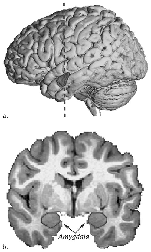
Figure 1.

Neuroanatomy of the human amygdala on MRI. A) Lateral view of a three-dimensional reconstruction of an MRI of a human brain with amygdala outlined (dashed line represents location of coronal slide in panel B and B) MRI coronal image with amygdala outlined.
