
Figure 1.
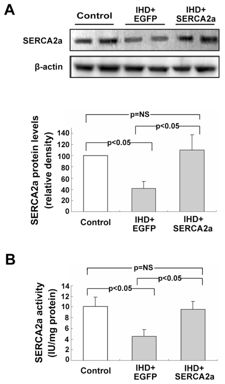
Myocardial expression and activity of SERCA2a protein after rAAV1-SERCA2a gene delivery. (A) Western blot and quantitative analysis of SERCA2a protein expression. β-Actin was used as an internal control. (B) Analysis of myocardial SERCA2a activity. Results show substantial decreases of SERCA2a protein and activity in the IHD+EGFP group compared with the control group, but activity was restored in the IHD+SERCA2a group. Data are presented as mean ± SEM (n = 4~6 per group).