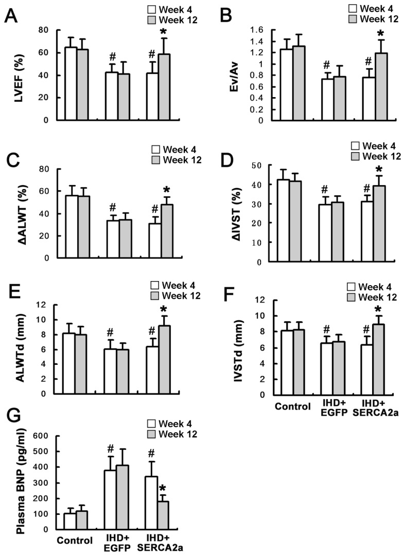
Figure 4.
Restoration of SERCA2a protein expression improves myocardial function. (A and B) Left ventricular ejection fraction (LVEF) and the ratio between early and atrial peak filling velocities (Ev/Av) showed that cardiac systolic and diastolic function was improved in the IHD+SERCA2a group 8 wks after viral administration. (C and D) Anterior lateral wall systolic thickening fraction (ΔALWT) and anterior lateral wall systolic thickening fraction (ΔIVST) showed the regional motion of anterior lateral and interventricular septal wall was improved in the IHD+SERCA2a group 8 wks after viral administration. (E and F) Anterior lateral wall diastolic thickness (ALWTd) and interventricular septal diastolic thickness (IVSTd) showed the thickness of anterior lateral and interventricular septal wall was increased in the IHD+SERCA2a group 8 wks after viral administration. (G) Serum BNP level was decreased in the IHD+SERCA2a group 8 wks after viral administration. Data are presented as mean ± SEM (n = 4~6 per group). #P < 0.05 versus control group; *P < 0.05 versus IHD+EGFP group.

