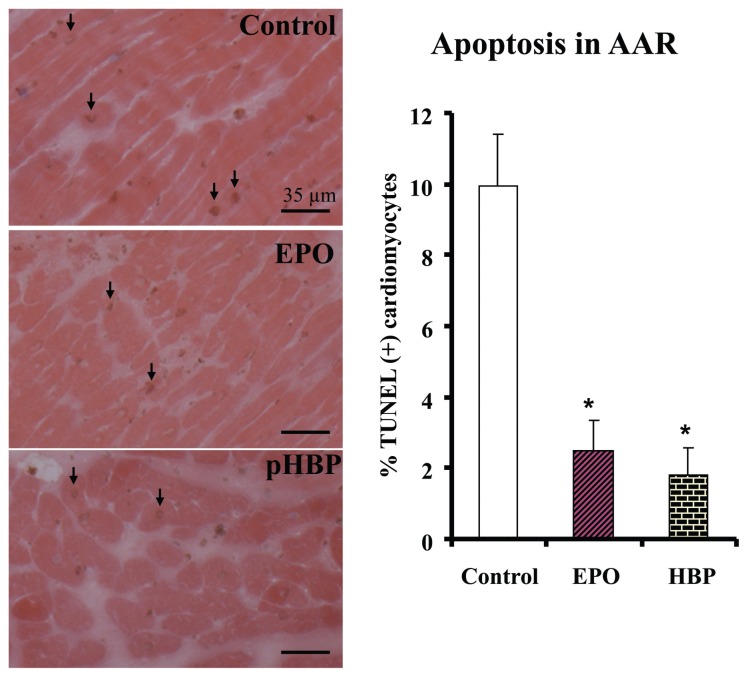
Figure 3.
TUNEL positive cardiomyocytes in the AAR of myocardium 24 h after coronary ligation and treatment with rhEPO or HBP. Left, representative examples of TUNEL staining in the AAR of myocardium. Right, percent of TUNEL positive cardiomyocytes in the AAR. The number of animals per group: Control, n = 8; rhEPO, n = 8; HBP, n = 9. P < 0.05 versus control (Bonferroni correction).

