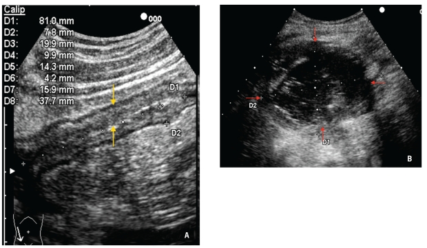
Figure 1.
Yellow arrows indicate bowel wall thickening with stenosis of the terminal ileum in a 30-year-old Crohn's disease patient (A). Ultrasonography of a superficial abscess (red arrows), which appears as a round hypoanechoic lesion with internal echoes and an irregular wall, in a 16-year-old female Crohn's disease patient (B).

