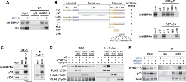
Figure 2.
MYBBP1A directly binds to p53, increasing the binding of p300 to p53. (A) Endogenous MYBBP1A associates with p53. MCF-7 cells were treated with siTIF-IA for 48 h. The cell lysates were immunoprecipitated with normal rabbit IgG or anti-MYBBP1A antibodies and analysed by immunoblot using antibodies against MYBBP1A and p53. (B) (Left) Domain structure of the full-length p53 and various deletion mutants. TAD, transactivation domain; PRD, proline-rich domain; DBD, DNA-binding domain; NLS, nuclear localization signal; TET, tetramerization domain; CRD, C-terminal regulatory domain; K, lysine residues that can be acetylated in the CRD region. (Right) MYBBP1A directly binds to the CRD region in p53. 35S-labelled MYBBP1A was incubated with the GST-fused full-length or truncated p53 proteins. After extensive washing, bound proteins were analysed by SDS–PAGE and autoradiography. (C) MYBBP1A forms a ternary complex with p53 and p300. MCF-7 cells were treated with siTIF-IA for 48 h. The cell lysates were sequentially immunoprecipitated with anti-MYBBP1A and anti-p300 antibodies, and immunoprecipitates were detected by immunoblot using antibodies against MYBBP1A, p53, and p300. (D) MYBBP1A increases the binding of p300, but not PCAF and Tip60, to p53. H1299 cells were transfected with expression vectors for FLAG-p300, FLAG-PCAF, FLAG-Tip60, MYBBP1A, and Myc-p53 as indicated. Twenty-four hours after transfection, the cells were treated with ADR (0.5 μg/ml) for 12 h to induce nucleolar disruption. FLAG-p300, FLAG-PCAF, and FLAG-Tip60 were immunoprecipitated using anti-FLAG antibodies, and the immunoprecipitates were eluted using FLAG peptides, then analysed by immunoblot using antibodies against FLAG and p53. HAT, histone acetyltransferases. (E) Knockdown of MYBBP1A decreases the binding of p300 to p53. MCF-7 cells were treated with siCont or siMYBBP with or without siTIF-IA for 48 h. The cell lysates were prepared, immunoprecipitated with normal rabbit IgG or anti-p300 antibodies, and analysed by immunoblot using antibodies against MYBBP1A, p53, and p300.

