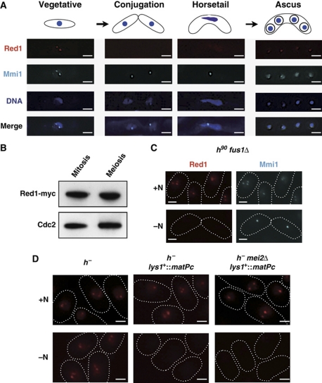
Figure 7.
Red1 foci are dynamic during meiosis. (A) Red1 signals disappeared at early in meiosis and then reappear in spores. Representative deconvolved images of a strain expressing both Red1-tdTomato and CFP-Mmi1 during meiosis are shown. DNA was stained with DAPI. Bars, 2 μm. (B) The protein level of Red1 does not change during meiosis. Cell lysates were prepared from a myc-tagged Red1-expressing strain in a vegetatively growing state or arrested at meiotic metaphase I and then analysed by western blotting. Cdc2 levels were also examined as a loading control. (C) Cell conjugation was not required for the loss of Red1 foci. fus1Δ cells expressing both Red1-tdTomato and CFP-Mmi1 were cultured with (+N) or without (−N) a nitrogen source at 26°C. Representative images are shown. The shapes of cells are indicated with white dotted lines. Bars, 2 μm. (D) Pheromone signaling but not nitrogen starvation triggers the disassembly of Red1 foci. h− strains carrying Red1-tdTomato, Red1-tdTomato/matPc, or Red1-tdTomato/matPc/mei2Δ were grown in the presence or absence of nitrogen for 12 h at 26°C. The white dotted lines indicate cell shape. Bars, 2 μm.

