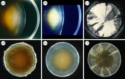
Figure 3.
(a,d) Brunescent nuclear cataract of moderate-to-marked density. The deep cortex is also coloured a yellowish brown. In (a) the broad light-scattering zone in the deep cortex is C3. Note that the cortical zone, C1, is intact. (b,e) A dense, non-brunescent, white nuclear cataract. Anterior and posterior subcapsular cataracts are also present. (a,b) Scheimpflug photography (courtesy of J. M. Sparrow) with the cornea seen to the right. (d,e) Dark-field micrographs of human donor lenses. (c,f) Spoke cataract of varying density and extent, viewed by retroillumination in a living patient (c) and dark-field illumination of an extracted lens (f); in neither case is there an associated nuclear cataract. Scheimpflug and retroillumination images (a–c) and dark-field micrographs (d–f) are from different subjects. Figure 3d reprinted from Michael et al. [64], © 2008, with permission from Elsevier.

