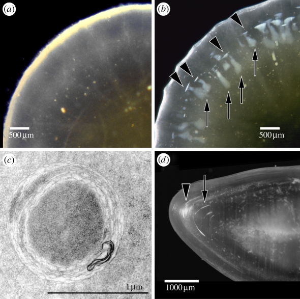
Figure 5.
Dark-field micrographs of aged human donor lenses, illustrating (a) small, dot-like opacities and (b) radial and circular shades. (c) Multilamellar body, as frequently found in human lenses with early cortical opacities probably causing the star-like opacities seen in (a). A slice cut in the axial plane of the fixed donor lens (b) is shown in (d). Arrows, radial shades and arrow heads, circular shades. Figure 5a,b reprinted from Michael et al. [64], © 2008, with permission from Elsevier. Figure 5c reprinted from Vrensen et al. [70], with permission from Taylor & Francis. Figure 5d reprinted from Michael [71], © 2010, with permission from Elsevier. Scale bars, (a,b) 500 µm, (c) 1 µm, (d) 1000 µm.

