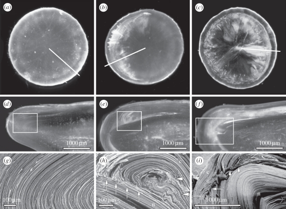
Figure 6.
(a–c) Panel of dark-field micrographs of aged human donor lenses and (d–f) of slices cut in the axial plane of the fixed donor lenses. The cutting plane is indicated by a white line in (a–c). Apart from some irregular scattering owing to imperfect slicing, the nuclear parts of the slices are free of opacification. In (b) a band-like opacity is seen. Except for the spokes in (c), the opacities are outside the pupillary space and will not have seriously influenced vision. Below is the fibre organization in a lens without cortical cataract and in two cases of cortical opacities (boxed areas in d–f), as visualized by scanning electron microscopy (g–i). (h) Fibres at the border zone between the C3 and C2 regions are broken (arrows) and the broken ends are directed towards the nuclear fibres, which maintain regular, uninterrupted organization. Further, note the curled (asterisk) and folded (arrowheads) fibres in the region adjoining the broken fibres. (i) The scanning electron microscopy micrograph shows broken fibres at several places (arrows) in the border zone between the C3 and C2 regions. The central fibres are regularly organized, as are the more superficial cortical fibres bridging the break zone. ep, epithelium; n, nuclear side; c, cortical side. Scale bars, (d–f,i) 1000 µm and (g,h) 100 µm. Figure 6 reprinted from Michael et al. [64], © 2008, with permission from Elsevier.

