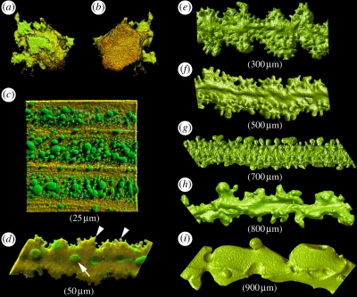
Figure 3.
Three-dimensional structure of mouse lens cells at various stages of differentiation as revealed by confocal microscopy. (a,b). Lens epithelial cells showing (a) basal or (b) apical surfaces. (c) Young elongating fibre cells located near the surface of a two-month-old mouse lens. The fibres are initially smooth and ribbon-like. Their membrane surface features a large number of gap junction plaques (green) visualized here by immunofluorescence with anti-connexin (Cx) 50. (d) At this stage, the fibre cell is in the process of losing its organelles. At the membrane surface, ball-and-socket processes (enriched with Cx50) are formed on the broad face of the lateral membrane (arrow). Smaller, finger-like structures protrude from the narrow membrane faces (arrowheads). (e) With the disappearance of organelles the fibres take on an undulating appearance. (f–i). Fibre cells dissected from progressively deeper cell layers. The primary fibre cells from the centre of the lens (i) are characterized by a very irregular structure. The approximate location (distance beneath the lens equatorial surface) of the fibre cells is indicated in parentheses. Mouse lenses of this age (two months) are approximately 1800 µm in diameter.

