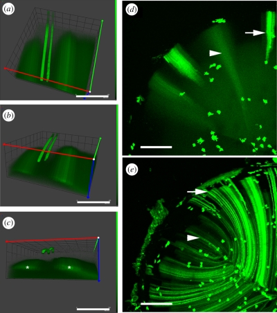
Figure 9.
Lim2-dependent cell fusion during mouse lens fibre cell differentiation. (a–c) A rotational series of volumetric reconstructions of a region of the outer cortex of a wild-type lens. GFP expression was induced in scattered lens cells. In the outer region (depths less than 50 µm) GFP is retained in the cytoplasm of expressing cells. Three such cells are shown. At greater depths, the cytoplasm of neighbouring cells becomes continuously linked through regions of partial membrane fusion. As a result, discretely labelled cells are not observed in the deeper cell layers. Rather, GFP diffuses among a cluster of neighbouring fibre cells (asterisks). The formation of cellular fusions is dependent on the presence of an intrinsic membrane protein, Lim2 (a.k.a. MP20). In wild-type mouse lenses (d) GFP is restricted to the cytoplasm of fibre cells located near the lens surface (arrow) but diffuses from expressing cells when those cells are buried to a depth of more than 50 µm (arrowhead). In the absence of Lim2 (e), fusions are not formed and GFP is retained by both superficial fibre cells (arrow) and fibre cells located deep below the lens surface (arrowhead). Scale bars, (a,b,c) 150 µm; (d,e) 250 µm.

