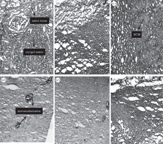
Figure 11.
Histological examination of the implanted constructs stained by H&E (a) after two weeks showing the integration of scaffolds with the native tissue and exhibiting the process of neo-vascularization (b) after four weeks showing the disintegration of the cryogel matrix (c) after six weeks showing the deposition of the ECM (d) control scaffold (scaffold implanted without chondorcytes) exhibiting the neo-vascularization process and infiltration of the cells from the native tissue (e) control scaffold after four weeks showing neo-vascularization (f) control scaffold after six weeks showing the disintegrated cryogel matrix and its integration with the native tissue.

