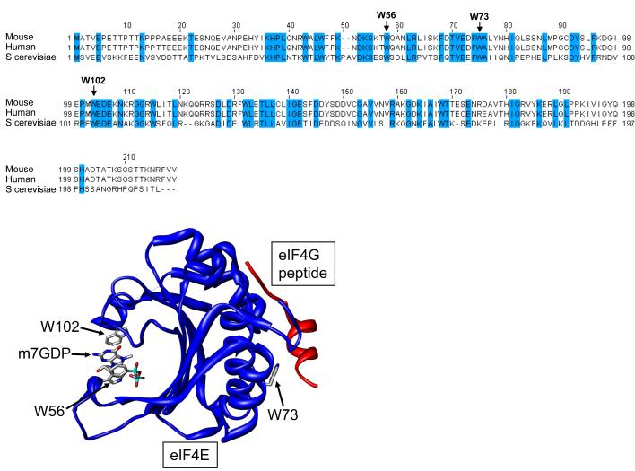
Figure 1. eIF4E sequence and structure.
Primary sequence of human (P06730), mouse (NM00917) and Saccharomyces cerevisiae (M15436) eIF4E, highlighting the high degree of sequence conservation. Three dimensional structure of mouse eIF4E in a complex with m7GDP and a peptide from eIF4G (PDB 1EJ4). Residues important for cap binding (W56 and W102) and ligand binding (W73) are highlighted

