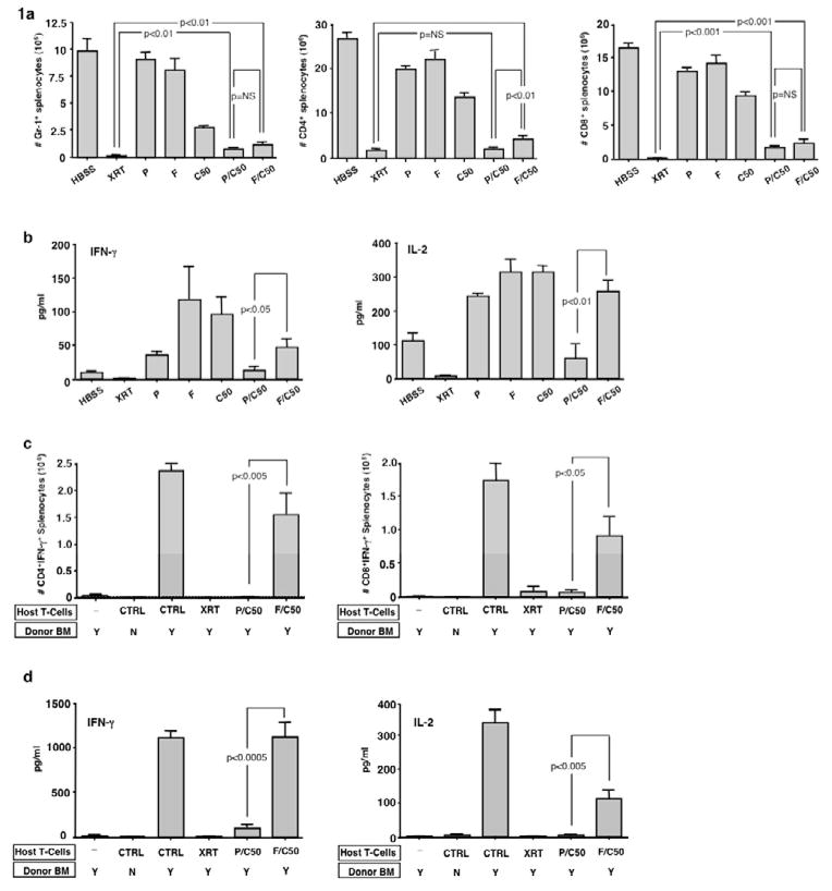
Figure 1. Pentostatin and cyclophosphamide synergistically induce host immune depletion and suppression.
BALB/c hosts were injected i.p. once daily for three days with either: HBSS (vehicle), pentostatin (P; 1mg/kg/d), fludarabine (F; 100mg/kg/d), cyclophosphamide (C50; 50 mg/kg/d), pentostatin plus cyclophosphamide (P/C50), fludarabine plus cyclophosphamide (F/C50) or treated with lethal total body irradiation (XRT; 950 cGy). (a) Three days post-therapy spleen cells were isolated, counted and analyzed by flow cytometry to determine the levels of depletion of myeloid cells (Gr-1+) and CD4+ and CD8+ T cells. (b) Spleen cells were co-stimulated and the resultant 24-hr supernatant was tested for IFN-γ and IL-2 content. (c) In a separate experiment, BALB/c hosts were lethally irradiated and then injected with syngeneic BALB/c host T cells (0.1 × 10⁶) obtained from mice treated with either HBSS (control), lethal irradiation (XRT; 1050 cGy), combination pentostatin plus cyclophosphamide (P/C50), or combination fludarabine plus cyclophosphamide (F/C50). One day following injection of host T cells, the hosts were transplanted with fully MHC-mismatched T-cell depleted bone marrow from B6 mice (10 × 106 cells). On day 8 post-BMT, spleen cells were isolated and stimulated with syngeneic or allogeneic dendritic cells for 24 hours. The number of host CD4+ and CD8+ cells producing allospecific IFN-γ was determined by cytokine capture flow cytometry. (d) Resultant culture supernatants were also tested for cytokine content (allogeneic stimulation condition is shown). All results are shown as mean ± SEM of n=5–10 per cohort.

