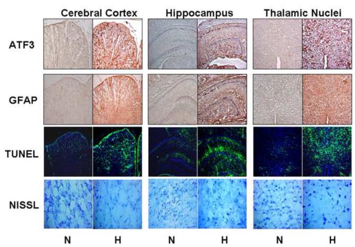
Figure 1. Chronic Fetal Hypoxia Results in Selective Anatomic Brain Injury.
Sample photomicrographs from mirror image coronal sections at the interaural level of 6.72mm to 5.40 mm (Bregma: from −2.28 mm to −3.60mm) including cerebral cortex, hippocampus, and thalamic nuclei from hypoxic (n=6) and control fetuses (n=6) stained for ATF3 (10x), GFAB (10x), TUNEL (10x), and Nissl (20x). Chronic hypoxia (10.5% O2 over the last 30% of gestation) visually increases ATF3, GFAB and TUNEL staining with an accompanying decrease in neuronal density.

