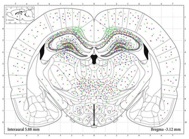
Figure 2. MAP of the Fetal Brain Injury Associated with Chronic Hypoxia.
Injured areas in the mirror sections were selected at the interaural level of 6.72mm to 5.40 mm (Bregma: from −2.28 mm to −3.60mm) were reconstructed based on quantification of immunostaining and TUNEL staining. The quantification of ATF3, GFAP and TUNEL were projected onto the standard coronal section. Chronic fetal hypoxia increased ATF3 (red), GFAP (green), and TUNEL (blue) staining specifically in cerebral cortex, hippocampus, and thalamic / hypothalamic nuclei. In cortex, the damage was widely distributed, while in hippocampus, the injury was greatest in cingulum, orices layer, and CA1, CA2, CA3 layers. In thalamic nuclei, the injury was greatest in the reuniens, central, ventral, and laterodorsal thalamic nuclei. The injury markers were also up regulated in the dorsomedial and ventromedial hypothalamic nuclei and lateral hypothalamus.

