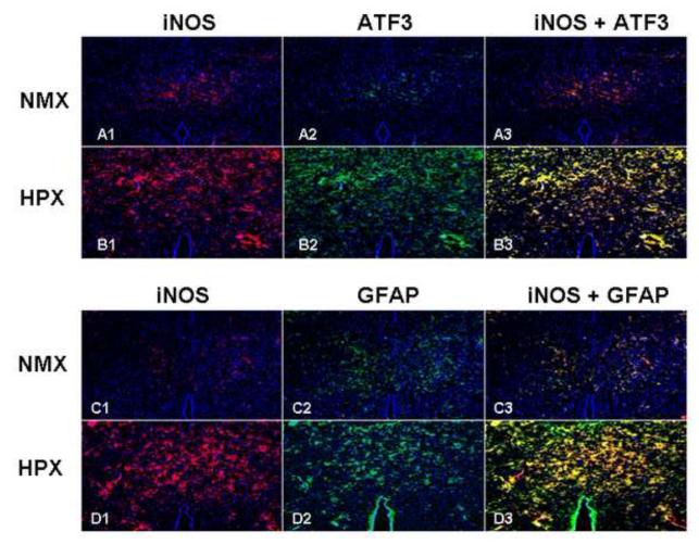
Figure 6. iNOS Co-localizes with Markers of Brain Injury.
Co-localization of iNOS and injury markers was sought using double fluorescent staining techniques (cross section hippocampus, ATF3 and GFAP, 10x). Chronic hypoxia increased iNOS (panels A1 vs. B1 and C1 vs. D1), ATF3 (panels A2 vs. B2) and GFAP panels (C2 vs. D2). iNOS co-localized with both ATF3 and GFAP positive cells (panels B3 and D3).

