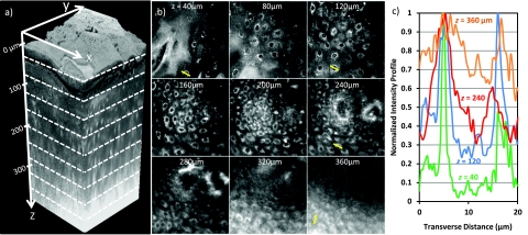
Figure 9.
(a) Three-dimensional rendering of a sequence of 200 lateral 2PAF images acquired from healthy human tongue biopsy. (b) Selection of lateral images from an imaging depth of 40 to 360 μm. Field of view in all lateral images is 170 μm. (c) Normalized signal profile from manually identified cells at imaging depths of 40, 120, 240, and 360 μm. Profiles are taken from lateral lines indicated in (b).

