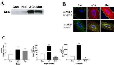
Fig. 1.
AC6 and AC6mut expression, localization, and activity. A, expression of AC6 and AC6mut proteins. Adult cardiac myocytes were incubated with Ad.AC6mut, Ad.AC6, or Ad.Null (control) for 40 h. AC6 and AC6mut proteins were detected by anti-AC5/6 antibody in immunoblotting. B, location of transgene proteins. Double immunofluorescence staining of AC6 and AC6mut by anti-AU1 antibody (red), anti-caveolin 3 (Cav-3) antibody (green), and anti-protein disulfide-isomerase (PDI) antibody (green). Hoechst dye was used to identify the nucleus (blue). There were no apparent group differences in cellular distribution: AC6 and AC6mut proteins were present in plasma membrane (associated with caveolin), nuclear envelope, and sarcoplasmic reticulum. C, AC activity. cAMP was measured in uninfected (Con) and Ad.AC6- and Ad.AC6mut-infected cardiac myocytes before (basal) and after 10-min stimulation with isoproterenol (Iso; 10 μM) or forskolin (Fsk; 10 μM). As expected, AC6 increased cAMP generation in response to isoproterenol and forskolin stimulation. AC6mut was associated with reduced cAMP generation in response to isoproterenol (a 59% reduction) and forskolin (an 80% reduction). Bars in the graphs denote mean ± S.D. (***, p < 0.001) derived from triplicates in three independent experiments.

