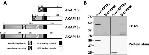
Fig. 3.
Direct binding of AKAP18γ and Inhibitor-1. A, schematic diagram depicting the known AKAP18 splice variants; PLN, phospholamban; LTCC, L-type calcium channel. B, GST-I-1 fusion protein (3 μg) was incubated with S-tagged AKAP18α, S-tagged AKAP18γ fusion protein (3 μg), or S-protein agarose alone. S-tagged pull-down assays were performed, and the association of GST-I-1 was determined by Western blot. Total protein was detected by Ponceau stain.

