Fig. 2.
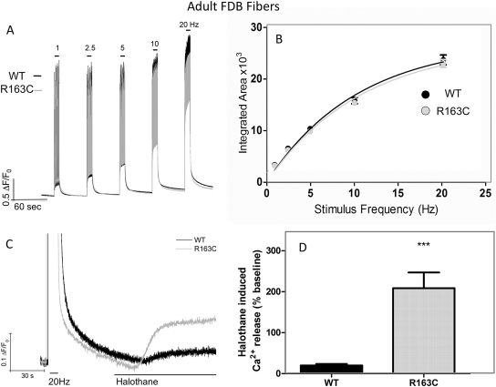
Adult FDB fibers isolated for R163C heterozygous MHS and WT mice do not differ in their responses to electrical stimuli. A, representative traces showing the frequency responses in WT (black traces) and R163C (gray traces) FDB fibers. Fibers were loaded with Fluo-4, and Ca2+ responses were elicited by electric field stimulation at 1, 2.5, 5, 10, and 20 Hz (10-s duration) as described under Materials and Methods. B, for each stimuli frequency, the integrated area was calculated and plotted as mean ± S.E.M. WT (n = 68) from four different fiber isolations and R163C (n = 78) from two different fiber isolations. C, representative trace showing that FDB fibers isolated from R163C show an amplified response to acute challenge with 0.1% halothane in the perfusion medium compared with FDB isolated from WT mice. D, amplitude of the Ca2+ response 30 s after the start of perfusion with halothane (0.1%). The magnitude of halothane-induced Ca2+ release was normalized to the baseline of each fiber 10 s before commencing perfusion of halothane. The data shown are mean ± S.E.M. obtained from n = 16 R163C and n = 24 WT fibers from two separate isolations (***, p < 0.001).

