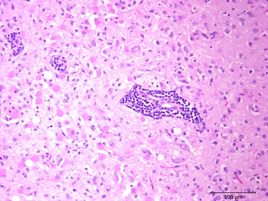
Figure 2.
This photomicrograph of cerebrum from the ferret shows a locally extensive area with large numbers of eosinophilic astrocytes (gemistocytic astrocytosis) and moderate numbers of small glial cells. Foci of blood vessels with mononuclear cell cuffing are visible also. Hematoxylin and eosin stain.

