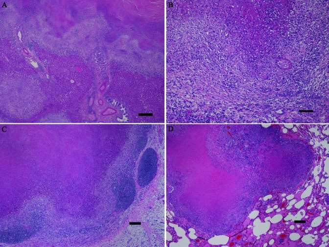
Figure 2.
Histologic findings. (A) This low-power photomicrograph of liver reveals extensive effacement of normal parenchyma by coalescing granulomas. (B) At higher magnification, the hepatic granulomas have central, massive necrosis, surrounded by intense granulomatous infiltrates including scattered giant cells, and with peripheral fibrosis. In the lower right portion of the image, biliary hyperplasia can be seen. (C) A mesenteric lymph node shows effacement of normal structure by a large granuloma. (D) One of the grossly identified nodules in the lung consists of coalescing granulomas. Note the lack of a thick, fibrous capsule in the lung granulomas, indicating more recent development. Hematoxylin and eosin stain; bar, 0.5 mm (A), 100 µm (B), 200 µm (C, D).

