
Figure 2.
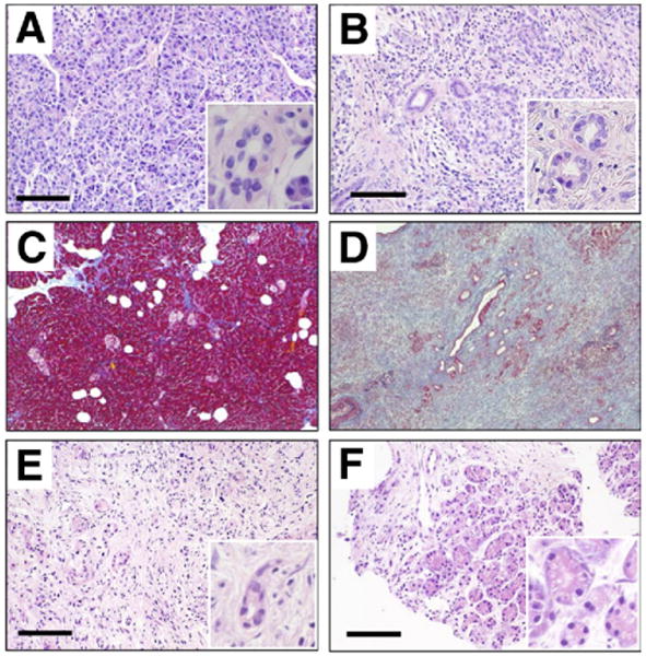
Pancreatic sections were stained with H&E. (A) Normal subject. (B) AIP, surgically resected tissue. Masson's trichrome staining of (C) normal pancreas and (D) AIP. H&E staining of specimen (E) before and (F) 3 months after initiation of steroid treatment. (E and F) Sections were obtained from the same patient. Bars, 100 μm. Insets show images at a higher magnification.