
Figure 5.
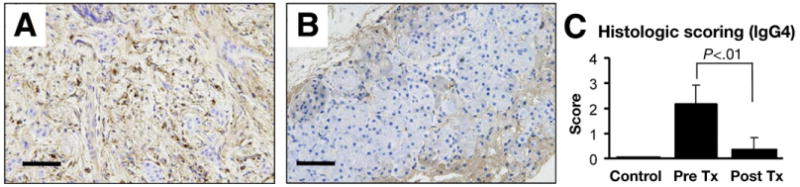
Immunohistochemical staining for IgG4. (A) Marked IgG4-positive plasma cell infiltrates are present in tissue before treatment. (B) The number of IgG4-positive plasma cells was decreased significantly after treatment. (C) Pancreatic sections obtained from normal subjects (n = 4), and AIP patients at 0 (n = 7) or 3 months (n = 7) treatment were scored at 0 (none), 1 (slight), 2 (moderate), and 3 (severe) for extent of IgG4-positive plasma cell infiltration.