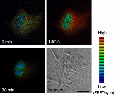
Fig. 3.
Ratio images of the cell co-expressing CFP-GR and YFP-importin α detected by FRET. COS-1 cells were co-expressed with CFP-GR and YFP-importin α and cultured in the absence of serum and steroids for at least 15 hr before observation. Fluorescent images of CFP-GR and YFP-importin α were captured using a filter set of CFP (440AF21 excitation, 480AF30 emission, and 455DRLP dichroic mirror) and YFP (500AF25 excitation, 545AF35 emission, and 525DRLB dichroic mirror), respectively. FRET image was detected using a filter set with 440AF21 excitation and 535AF26 emission, and 455DRPL dichroic mirror at 0, 10, and 30 min after treatment with 10−6 M CORT. Filter sets were purchased from Omega Optical Inc. The ratio of the FRET image was divided by donor image to obtain the ratio images using MetaMorph software (Universal Imaging Corp.). The ratio images were pseudo colored. The red range showed high ratio and blue range showed low ratio. High ratio was observed in the cytoplasm, indicating an interaction of CFP-GR and YFP-importin, whereas low ratio was observed in the nucleus, indicating a dissociation of these two molecules.

