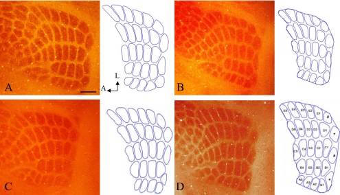
Fig. 1.
Cortical barrel field, including the PMBSF. Twenty-seven barrel areas of PMBSF were outlined for measurement using ImageJ. Control group (A, B), BPA-exposure group (C, D), males (A, C) and females (B, D). No significant differences were shown in the number and patterning of PMBSF between treatments and sex. COX staining. Bar=300 µm.

